Vision in Dogs and Cats
Do animals see in black and white? Answers to common questions from clients.


Figure 1. Color vision in a human (left) 3° and a dog (right). Companion animals do not distinguish between red, green, and yellow. (Image from Cook1; used with permission)
Every now and then veterinarians are asked about animal vision: “Are dogs truly color blind?” or “Do cats have superior night vision?” or “Who sees better, my dog or my cat?” Vision is an extremely complex sense that is influenced by various elements, differs significantly among species, and can be tested using many methodologies. Thus, there is no easy way to answer these questions.
It’s true that some animals, including cats and dogs, may be partially color blind, but certain aspects of their vision are superior to that of humans. Not only is it interesting to imagine how your patients see the world, but having a better understanding of their vision can positively influence, for example, the choice of lighting you use in your office.
RELATED:
- Prevalence of Ophthalmologic Diseases in Other Countries
- Dry Eye in Dogs
Although this review is not intended to offer an exhaustive discussion of the topic, it does focus (pun intended) on select aspects of vision in our patients.
Do Your Patients See in Black and White?
We’ll start with the most popular question: Are cats and dogs able to see color? Cone photoreceptors are responsible for color vision in all species. The richness of color vision, and the number of shades that an eye can see, are determined by the number of cone photopigment populations and the degree of overlap in their absorbance spectrum.
To better understand how your patients see, it’s helpful to know that humans have 3 cone photopigment populations, with peak absorbance in the red, blue, and green wavelengths—which is why these are our 3 primary colors—making humans a trichromatic species. On the other end of the spectrum are monochromatic species, which have only a single photopigment population. This type of vision is characteristic of nocturnal species, which can perceive only shades of a single photopigment, usually with a red/green hue.
Most domestic animals are dichromatic, meaning they possess 2 photopigment populations. So, contrary to popular belief, domestic animals do not see in black and white. They have color vision, although not as rich as that of us trichromatic humans.
Cats, dogs, cattle, and horses all have 2 photopigment populations, with absorbance peaks of about 440 and 555 nm. Consequently, these species can see blue but have difficulty distinguishing between red, green, and yellow. Color vision in these species is analogous to that of “color-blind” humans, who are usually missing their red and green cone population (Figure 1).1
If you think humans have the most superior vision, however, you’d be mistaken. Many fish and bird species are tetrachromatic, meaning they have a fourth photopigment absorbing light in the ultraviolet end of the color spectrum. As such, they possess color vision that is significantly richer than humans.
Visual Fields
The frontal part of the visual field is covered by both eyes, resulting in binocular vision, which is traditionally required for depth perception. The monocular visual field is the lateral visual field of each eye, in which there is no input from the other eye. The extent of the monocular and binocular visual fields is largely dictated by the location of the orbits and eyes in the skull.


Figure 2. The extent of the visual field in a dog, horse, and cat. Note that the panoramic vision of the horse comes at the expense of a small, frontal binocular visual field. Predators such as dogs and cats have a large frontal, binocular field of vision essential for depth perception, but suffer from a large blind spot behind them (Courtesy: Mr. John Doval, UC Davis).
Species with front-facing eyes have a large binocular visual field. Consequently, they have 2 small lateral monocular visual fields and a large posterior blind spot. Frontal eyes are found in predator species that require superior depth perception for accurate spatial localization when pouncing on their prey. Dogs, cats, and raptor birds fall into this category. The evolutionary price paid in the form of small peripheral fields and a large blind spot is of less concern to predator species.
On the other hand, herbivores have lateral orbits, giving them a small frontal binocular visual field and 2 large lateral monocular fields of vision that give them almost 360° panoramic vision. The large lateral monocular fields are important to these species as most of them need to see approaching predators.
How do visual fields compare? The total visual field of cats is estimated at 200°, including a 140° frontal binocular field and 2 lateral monocular fields of 30° each (Figure 2). Horses are estimated to have a visual field of about 355°, including a frontal binocular field of 65° and 2 large lateral monocular fields of about 145° each. To know the implication of these numbers, consider that enucleation of 1 eye would cause a cat to lose 30° of its total visual field and a horse nearly 145° of its total visual field. With the loss of an eye, then, a horse could become dangerous and easily scared by objects moving on the side with vision loss.
Pets and TV
When photoreceptors are stimulated by a flash of light, they need a minimum amount of time to recover before they can respond to a subsequent flash. If the flashes are presented rapidly, the photoreceptors do not have enough time to recover fully. Beyond a certain threshold, flashes become so rapid that the retina is unable to recover. At this point, the retina is no longer able to perceive or respond to the individual flashes, resulting in “fusion” of the responses and the perception of a continuous, rather than a flickering, light.
In humans, cone responses fuse at 45 Hz. Therefore, images projected on television or computer screens, which flicker at 60 Hz (in the United States), are perceived as a continuous image. But in many other species, cone responses fuse at 70 to 80 Hz. As a result, pets can perceive individual flickering images when watching television, which can have a dramatic effect on their interest in the program.
For the same reason, companion animals can detect the flickering of fluorescent lights. This is an important distinction that should be considered when selecting lighting for your hospital. Yellow, incandescent or LED bulbs that do not flicker provide the patient with a more hospitable atmosphere.


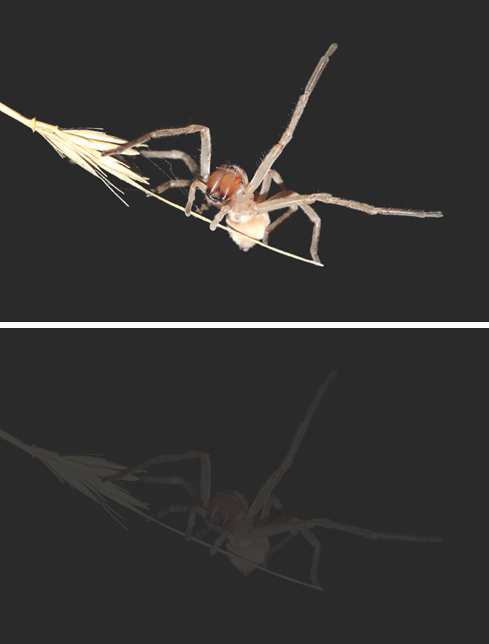
Figure 3. The same insect viewed by a cat (top) and a human (bottom). At night, a cat’s vision is about 6 times better than a human’s. (Courtesy: Shlomi Levi, DVM, Hachaklait)
Night Vision
The common narrative that companion animals have very sensitive night vision is based in truth. Study results show that cats can detect light that is 6 times dimmer than the lowest detectable threshold of humans (Figure 3).2
This is due to several factors. First is the amount of light entering the eye. Cats have very large corneas and pupils, with diameters about 50% larger than those of humans. Therefore, more light can pass through feline eyes and reach the retina.
Second, with the exception of pigs, all domestic species possess a tapetum lucidum, a reflective tissue located in the choroid behind the retina. Light that is not absorbed by the retinal photopigment strikes the tapetum and reflects back to the retina, doubling the chances that it will be absorbed. Thus, the tapetum acts as a mirror that helps the animal increase retinal illumination in dim light. But there is an evolutionary price to be paid. The light reflected by the tapetum is scattered in the eye, reducing visual resolution. This has little consequence at night when cones, which are responsible for high-resolution vision, are inactive, but it becomes very detrimental in daylight.
The third and most critical factor that affects night vision is the distribution and concentration of retinal photoreceptors. In the retinal periphery, the maximal rod concentration in cats is about 5 times higher than that in humans. Because rods are much more sensitive to low light levels, and in fact can detect a single photon, this high rod concentration provides cats with their highly sensitive night vision and motion perception.
Visual Resolution and Acuity
Visual resolution is determined mostly by the optics of the eye and the anatomy of the retina. Optically, in an emmetropic eye, light focuses properly on the retinal photoreceptors, contributing to a sharp image. In a short-sighted (myopic) eye, the image is focused in front of the retina, while in a far-sighted (hyperopic) eye the image is focused behind the retina, both of which result in a blurry image.
Findings from a study of 1500 dogs showed that a majority are emmetropic, meaning the optics of their eyes are calibrated to generate a focused image on the retina.3 If these dogs were humans, they would not require corrective glasses. The same study’s results showed that about 25% of all dogs are short-sighted, with a refractive error ranging from —0.5 to –6.0 D.
For some dog breeds, the average refractive error is myopic. For example, two-thirds of Rottweilers studied were short sighted, with a mean refractive error of —1.8 D.3,4 Similarly, significant numbers of dogs were far sighted, with certain breeds having a mean far-sighted refractive state.7 For example, the Australian shepherd has a mean refractive error of +1.3 D.
Visual resolution is also determined by retinal anatomy. As noted, factors that contribute to enhanced night vision have a detrimental effect on daytime vision and visual resolution. Thus, the presence of a tapetum lucidum in the eyes of dogs and cats causes scattering of light that reduces visual resolution. Furthermore, differences in the concentration of cones, which process high-resolution images, also affect visual resolution. In companion animals, the concentration of these cells is lower than in humans, resulting in lower visual resolution. For example, the maximal cone concentration in the feline retina is one-eighth that in the human retina. Consequently, even if a dog or a cat is emmetropic, and has a well-focused image on the retina, its visual resolution will be reduced because the resolving power of the eye is attenuated by the low cone concentration in the retinas.
Visual acuity is typically expressed as a Snellen fraction, and this is where people have the advantage. The acuity of normal humans is the well-known figure, 20/20. By comparison, the visual acuity of horses is estimated to be 20/33, meaning that a horse must be 20 feet from an object to see it as well as a person standing 33 feet away can see it.
Visual acuity is 20/75 in dogs and 20/150 in cats. In other words, a cat has half the acuity of a dog and a fifth of the acuity of a horse. The simple translation: A cat has to be more than 7 times closer to an object to see it as sharply as we do! These figures are based on the assumption that the animal is emmetropic. If the animal is not emmetropic, then its visual resolution will be poorer.
It all seems so clear now, doesn’t it?
Dr. Ofri is a professor of comparative ophthalmology at the Koret School of Veterinary Medicine, Hebrew University of Jerusalem in Rehovot, Israel.
References:
- Cook C. What do they see and how do we know? Clean Run. 2009;Jan:61-66.
- Weale RA. The spectral reflectivity of the cat's tapetum measured in situ. J Physiol 1953;119(1):30-42.
- Kubai MA, Bentley E, Miller PE, Mutti DO, Murphy CJ. Refractive states of eyes and association between ametropia and breed in dogs. Am J Vet Res. 2008;69(7):946-951. doi: 10.2460/ajvr.69.7.946.
- Hernandez J, Moore C, Si X, Richer S, Jackson J, Wang W. Aging dogs manifest myopia as measured by autorefractor. PLoS ONE. 2016;11: e0148436. doi: 10.1371/journal.pone.0148436.
Suggested Reading:
- Imamoto Y, Shichida Y. Cone visual pigments. Biochim Biophys Acta. 2014;1837(5):664-673. doi: 10.1016/j.bbabio.2013.08.009.
- Miller PE. The eye and vision. In: Maggs DJ, Miller PE, Ofri R (Eds). Slatter’s Fundamentals of Veterinary Ophthalmology, 6th ed. St Louis, MO: Saunders Elsevier 2018:1-17.
- Miller PE, Murphy CJ. Equine vision: normal and abnormal. In: Gilger BC (Ed). Equine Ophthalmology. 3rd ed. Ames, IA: John Wiley & Sons; 2017:508-544.
- Miller PE, Murphy CJ. Vision in dogs. J Am Vet Med Assoc. 1995;207(12):1623-1634.
- Neitz J, Geist T, Jacobs GH. Color vision in the dog. Vis Neurosci. 1989;3(2):119-125.
- Ofri R. Optics and physiology of vision. In: Gelatt KN, Gilger BC, Kern TJ (Eds). Veterinary Ophthalmology. 5th ed. Ames IA: John Wiley & Sons; 2013:304-410.
- Ofri R, Hollingsworth SR, Groth A, et al. Effect of optical defocus on performance of dogs involved in field trial competition. Am J Vet Res. 2012;73(4):546-550. doi: 10.2460/ajvr.73.4.546.


UN, WHO address public health concern over avian flu transmission to humans
April 18th 2024Veterinary professionals working with certain animals are advised to take precautionary steps to minimize risk of infection, while researchers in Texas study potential H5N1 vaccines, antivirals, and antibody therapies for humans
Read More

