Disease State Watch: Orthopedics
The latest research on treating orthopedic conditions.


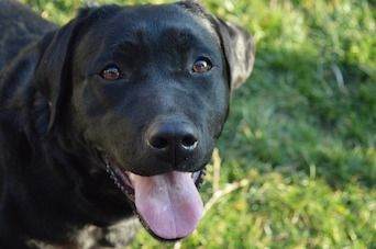
Tibial Plateau Leveling Osteotomy (TPLO) is the preferred surgical procedure for treating cranial cruciate ligament rupture, but osteoarthritis (OA) remains a risk.
To see if a daily joint supplement could lead to better outcomes, investigators in Italy combined TPLO with a therapy containing chondroitin sulfate, glucosamine hydrochloride, N-palmitoyl-D-glucosamine, quercetin, vitamin E, and omega-3 fatty acids. The controlled, open-label, preliminary study enrolled 13 client-owned dogs recently diagnosed with cruciate ligament rupture. All the dogs underwent TPLO surgery and were then randomized to 2 groups. The treatment group received a daily joint supplement throughout the 90-day study; the control group did not.
Synovial fluid samples were collected before surgery and at 30 and 90 days after. Using high-resolution proton nuclear magnetic resonance spectroscopy, a technician blinded to the treatment group analyzed the samples for lactate and creatine, both of which increase in OA. At 30 days, lactate and creatine concentrations were higher for dogs in the control versus the treatment group, but no significant difference was noted at 90 days.
The dogs were examined for lameness at the same time intervals. Radiographs were also taken, and an OA score was assigned. Although lameness scores improved for both groups, there were no significant differences in OA scores, lameness scores, or radiographic changes. However, because inflammation likely plays a role in the development of OA, a joint supplement with antioxidant, anti-inflammatory, and analgesic effects may help by limiting the subsequently damaging inflammatory response, according to the investigators.
Reference:
- Martini FM, De Bellesini AB, Miolo A, Del Coco L, Fanizzi FP, Crovace A. Combining a joint health supplement with tibial plateau leveling osteotomy in dogs with cranial cruciate ligament rupture. An exploratory controlled trial [published online October 6, 2017]. Int J Vet Sci Med. doi: 10.1016/j.ijvsm.2017.09.006.
Unique Canine ACL Reconstruction Procedure Shows Promise for Clinical Use
Investigators exploring improved grafts for use with anterior cruciate ligament (ACL) reconstruction, a common procedure, have developed a novel “hybrid double-bundle” canine model. This unique configuration of a quadriceps tendon allograft with internal brace (QTIB) allows for an effective biologic-synthetic load-sharing ACL construct.
The University of Missouri team surgically transected the right ACLs from 10 purpose-bred research hounds and used the QTIB construct with suspensory fixation to reconstruct the ACL apparatus. The dogs’ left knees remained unaltered and formed the nonsurgical control group.
Postoperative radiographs were used to assess the surgical sites and implant placement. The dogs were given analgesics for at least 3 days postoperatively and were restricted to their kennels during the study, let out only for monitored exercise. The limbs that underwent surgery were maintained in soft padded bandages for 2 weeks.
At 2, 3, and 6 months postoperatively, the dogs were examined by a board-certified orthopedic surgeon who was blinded to treatment group. Both knees of each dog were palpated, and anterior drawer and degree of internal rotation were measured. Each dog was also assessed for lameness, pain, effusion, and range of motion in the knees.
At 2 and 6 months, radiographs were repeated, and each knee that had undergone surgery was assessed arthroscopically for evidence of osteoarthritis (OA) and to reassess the surgical sites and implanted apparatus. At 6 months, the dogs were euthanized humanely, and tissues in and around the knees were evaluated.
The investigators reported that all dogs could bear weight within 48 hours after surgery and remained functional at each assessment. The reconstructed knees stayed clinically stable, and differences in cranial drawer and internal rotation were not significantly different between groups. Further, there were no radiographic changes consistent with OA for 6 months postoperatively. According to the investigators, these results indicate that a QTIB used for complete ACL reconstruction would provide sustained knee function without the development of premature OA, a concern with prior models.
Reference:
- Cook JL, Smith P, Stannard JP, et al. A canine arthroscopic anterior cruciate ligament reconstruction model for study of synthetic augmentation of tendon allografts. J Knee Surg. 2017;39(7):704-711. doi: 10.1055/s-0036-1597618.
Promising New Treatment Option for Canine OA Induced by Hip Dysplasia
Hip dysplasia (HD) can progress to hip osteoarthritis (OA) in dogs, resulting in chronic pain and reduced mobility. Nonsteroidal anti-inflammatory drugs and nutraceuticals (commonly containing glucosamine hydrochloride and chondroitin sulfate) are considered traditional therapies, but a potential treatment for humans with OA might help dogs, too. A 2016 study compared the efficacy of intra-articular (IA) injections of hyaluronic acid—a naturally occurring glycosaminoglycan component of cartilage matrix and synovial fluid—with traditional treatment in a group of client-owned dogs with OA induced by hip dysplasia.
Investigators from the Oeste Paulista University in São Paul, Brazil, enrolled 16 client-owned dogs with HD and OA into a randomized, controlled, 90-day study. The treatment group received IA hyaluronic acid injections; the control group received IA injections of saline solution as well as an oral nutraceutical (containing glucosamine, chondroitin, and collagen) and carprofen.
At the beginning of the study, a veterinarian blinded to treatment group assigned scores for pain, lameness, and ability to jump and climb stairs. Additional veterinary and pet-owner evaluations were performed prior to treatment and at 15, 30, 60, and 90 days after the injections, using the Helsinki Chronic Pain Index and the Canine Brief Pain Inventory (CBPI).
Both groups improved significantly over 90 days compared with baseline, although dogs in the treatment group received better CBPI scores at 60 to 90 days compared with controls. Further, half the dogs in the control group and all the dogs in the treatment group improved clinically based on total CBPI evaluation.
Pet owner assessments using CBPI indicated an average clinical improvement of 56.4% in treatment group dogs and 29.6% in control group dogs, a statistically significant difference. At the end of the treatment period, 75% of pet owners in the treatment group categorized their dog’s quality of life (QoL) as “very good” to “excellent,” and 75% of owners in the control group rated their dog’s QoL as “good.”
The investigators concluded that IA hyaluronic acid injections may represent a viable treatment for dogs with hip OA, but long-term studies are needed.
Reference:
- Carapeba GOL, Cavaleti P, Nicácio GM, Brinholi RB, Giuffrida R, Cassu RN. Intra-articular hyaluronic acid compared to traditional conservative treatment in dogs with osteoarthritis associated with hip dysplasia. Evid Based Complement Alternat Med. 2016;2016:2076921. doi: 10.1155/2016/2076921.