Snakebite in Animals: A Brief Refresher
Each year, 7000 to 8000 people1 and an estimated 150,000 dogs and cats2 in the United States are bitten by venomous snakes. Venomous snakes native to North America belong to 2 groups: crotalids and elapids. Crotalids, or pit vipers, include rattlesnakes (found throughout the United States), copperheads (in eastern states), and cottonmouths or water moccasins (in wetlands of the southeastern United States). The only elapid in the United States is the coral snake, which lives in wooded or marshy areas in the South.1
Crotalid bites occur much more often than elapid bites.3 Bites from venomous snakes not native to the United States have also been reported in humans, including children; these bites are mostly by snakes kept in residences (not zoos).4 It is reasonable to assume that pets could also be bitten by non-native snakes for which antivenin is not readily available.
Fatalities are more common in dogs than in other animals,3 occurring in an estimated 1% to 30% of cases.5 Mortality after snakebite is low in cattle and horses because of their size, although bites to the head can result in swelling that leads to asphyxiation and death. Not all bites result in envenomation, however. Many are “dry bites,” with little or no venom delivered.3
First Aid
Snakebite is a medical emergency. Treatment at a veterinary hospital should not be delayed by attempts to provide first aid.3 First aid should consist of limiting the animal’s activity and transporting it as quickly as possible. Most field treatments are ineffective at best, and some can harm the patient. Cold packs, hot packs, incision and suction, and tourniquets should not be used.3,5 Because elapid venom can cause respiratory paralysis, animals bitten by coral snakes should be taken to a facility that can provide 24-hour monitoring and mechanical ventilation.6
Crotalid Bites


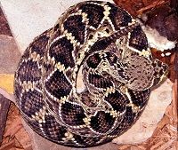
Crotalus adamanteus, “Eastern diamondback” rattlesnake
Crotalid bites typically cause local tissue necrosis with swelling, discoloration, and epidermal sloughing.3 Local reactions are generally more severe with rattlesnake bites than with copperhead or cottonmouth bites. Blood or serum may seep from bite wounds. However, fang marks do not necessarily indicate envenomation. A tissue reaction is usually seen within 10 minutes of moderate to severe envenomation. If no swelling has occurred within 1 hour and no systemic signs of envenomation are present, the bite was probably dry.5 Systemic clinical signs include coagulopathy, petechiae, weakness, vomiting, diarrhea, hypotension, thrombocytopenia, pulmonary edema, cardiac arrhythmia, and shock.2,5


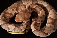
Agkistrodon contortrix “Southern copperhead” snake
The venom of Mojave, timber, and canebrake rattlesnakes is primarily neurotoxic. Envenomation causes neurologic signs such as muscle fasciculations.5 It is possible for an animal to die from neurotoxic crotalid venom without showing any local tissue reaction.2,5


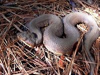
Agkistrodon piscivorus leucostoma, “Western cottonmouth” snake
If a crotalid bite is not witnessed, it can be mistaken for other disorders. Differential diagnoses include trauma, abscess, bite wound from an animal other than a snake, spider bite, and allergic reaction to an insect bite.3,5
Elapid Bites


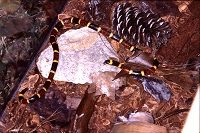
Micrurus tener, “Texas coral snake"
The predominant signs of coral snake envenomation are neurologic, with minimal local swelling and pain. The onset of signs may be delayed for up to 18 hours. Signs include tetraparesis, weakness, ataxia, muscle fasciculations, tachypnea, shallow breathing, altered (quiet) mental state, hemolysis, anemia, and hemoglobinuria.3,6 Death, when it occurs, is usually caused by respiratory paralysis.5
Puncture wounds from coral snakes may be difficult to find. Differential diagnoses for elapid envenomation include tick paralysis, botulism, and myasthenia gravis.5
Diagnostic Tests
Diagnostic testing includes a complete blood count, coagulation profile, chemistry panel, and urinalysis. Anemia and thrombocytopenia can occur with both crotalid and elapid bites, and echinocytes may appear after rattlesnake and coral snake envenomation. Coagulation deficits may or may not be accompanied by thrombocytopenia. Rhabdomyolysis can cause an increase in serum creatine phosphokinase. Urinalysis may reveal hematuria, hemoglobinuria, or myoglobinuria. Pigmenturia indicates severe envenomation. Using a snakebite severity score is recommended to track the progression of signs and help decide whether antivenin is needed.5
Treatment
Snake envenomation patients should be observed for at least 8 hours.5 Treatment for crotalid envenomation can include intravenous crystalloids, opioid analgesics, and treatment of coagulopathy. Rapid-acting corticosteroids, antibiotics, and antivenin are used in some cases.3 The use of steroids is controversial, and nonsteroidal anti-inflammatory drugs should not be used if coagulopathy is present. Antivenin may be indicated if swelling progresses rapidly, if coagulopathy or thrombocytopenia is present, or if the patient has signs of neuromuscular toxicity or shock. Antivenin is often not needed for copperhead bites. Newer Fab antivenins have a lower risk of anaphylaxis but could be too expensive for veterinary patients.5
Treatment for coral snake envenomation consists of intravenous fluids, anticonvulsants, and respiratory support.3 Patients should be monitored in the hospital for 48 hours, and mechanical ventilation may be needed for up to 72 hours.6 Coral snake antivenin is not currently available in the United States.3
Prognosis
The prognosis for snakebite patients depends on the type of snake, degree of envenomation, size of the patient, location of the bite, and time before initiation of treatment. Severe crotalid envenomation can cause significant long-term effects secondary to tissue necrosis3; limb edema may persist for months.5 Elapid bites usually do not cause significant long-term sequelae in patients that survive.3
Dr. Laurie Anne Walden received her doctorate in veterinary medicine from North Carolina State University in 1994. After an internship at Auburn University College of Veterinary Medicine, she returned to North Carolina, where she has been in companion animal general practice for over 20 years. Dr. Walden is also a board-certified Editor in the Life Sciences and owner of Walden Medical Writing.
References
- Venomous snakes. Centers for Disease Control and Prevention website. www.cdc.gov/niosh/topics/snakes/. Updated July 24, 2015. Accessed January 5, 2016.
- Peterson ME. Snake bite: pit vipers. Clin Tech Small Anim Pract. 2006;21(4):174-182.
- Overview of snakebite. Merck Veterinary Manual website. www.merckvetmanual.com/mvm/toxicology/snakebite/overview_of_snakebite.html. Reviewed/revised July 2013. Accessed January 5, 2016.
- Warrick BJ, Boyer LV, Seifert SA. Non-native (exotic) snake envenomations in the U.S., 2005—2011. Toxins (Basel). 2014;6(10):2899-2911. doi: 10.3390/toxins6102899.
- Gilliam LL, Brunker J. North American snake envenomation in the dog and cat. Vet Clin North Am Small Anim Pract. 2011;41(6):1239-1259. doi: 10.1016/j.cvsm.2011.08.008.
- Peterson ME. Snake bite: coral snakes. Clin Tech Small Anim Pract. 2006;21(4):183-186.